Codec 5

Friday, September 26, 2008

Week 14

Lab Technique: Radioimmunoassay

It’s the 14th week already. 6 more weeks to go. Hope you guys are doing fine. =)

Last week I mentioned about faecal extraction. So now, after the extraction has been done, we can now measure the glucocorticoid level.

To measure the glucocorticoid levels in the faecal sample, radioimmunoassay (RIA) is used. Just to refresh your memory. The principle of RIA is based on the competition between unlabelled ligand and a fixed amount of radiolabelled ligand for a limit amount of antibody. The proportion of the bound labelled ligand is inversely related to the concentration of the unlabelled ligand.

An essential step in RIA is the separation of the unbound radiolabelled ligand from the Ab-Ag complexes. This is done so that the unbound radiolabelled ligand will not interfere with the counting and hence cause inaccurate result. However, this step can make the whole RIA procedure to be time consuming and can be prone to human error due to additional steps needed as compared to using SPA.

What is SPA?

SPA refers to scintillation proximity assay that makes use of SPA reagent to eliminate the separation step. The SPA reagent contains either a secondary antibody or protein A that is bound to a fluomicrosphere. The fluomicrosphere will produce light if it is bound to a radiolabelled ligand. Light will not be produced if the SPA reagent is bound onto an unlabelled ligand. Unbound radiolabelled ligand will also not produce light.
F = Fluomicrosphere with protein A or secondary Ab attached
Ab = Primary Ab
L = Unlabelled Ligand
L* = Radiolabelled ligand

To calculate the concentration of the unlabelled ligand, a machine called the scintillation counter is used. Depending on what kind of isotope used, the scintillation counter can vary. If a beta emitting isotope is used, a beta-scintillation counter will be used and if a gamma emitting isotope is used, a gamma-scintillation counter will be used. The scintillation counter will calculate the amount of radiolabelled ligand by detecing the light and then using a software, the concentration of unlabelled ligand can be determine by interpolating from a standard curve. By detecting only the light produced by bound radiolabelled ligand, this eliminate the need for the separation step.

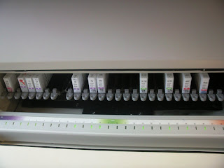
This is a beta-scintillation counter.
(Wallac 1414 Liquid Scintillation Counter)
In our lab, tritium (3H) is used. And since tritium is a beta emitting isotope, a beta-scintillation counter will be used. FYI, handling of any radioisotope compound has to be done in a radioactive room. The radioactive room looks very much like a normal lab. It is just that in front of each bench, there would be a shield and we are expected to work behind the shield for safety reason.

Again, depending on whether a beta emitting radioisotope is used or a gamma emitting isotope is used, the shield could vary. Beta emitting isotopes are weakly penetrative, hence, a plastic shield is sufficient enough to block the rays. In fact, a piece of paper is already enough to block off the beta-rays. Gamma emitting isotope such as iodine 125 is more penetrative. Hence, when using 125I, we are expected to work behind a metal shield. Also, as the rays emitted can be absorbed through skin, gloves as to be wore at all times!

And of course, we will have controls to ensure that our results are valid.

As the steps involved in carrying out the RIA is quite long, I will just give a brief outline on what is done. When measuring different ligands, different set of standard concentration, antibody and tracer will be used. Hence, for illustration, I will focus on cortisol.

First we have to prepare our standards so that a standard curve can be plotted.

6 standards are prepared with concentration consisting of 188, 375, 750, 1500, 3000 and 6000 fmol/0.1ml by serial dilution starting from the standard of the highest concentration. FYI, the standard with the highest concentration is already pre-prepared by the staff in the lab.

After which, we need to prepare the SPA reagent and antibody mixture and also the 3H cortisol tracer.

Because the controls used are in serum, there is also a need for the extraction of steroids as the steroids will be bound to its binding globulin. In this case, we don’t extract the steroids like what was done on the faeces. The controls are first diluted in distilled water and then heated at 60oC for 30 minutes. The heating is sufficient to dissociate the steroids from its binding globulin.

Next, we will dilute the faecal samples 50x. After which, 100 µl of standards, PBS, faecal samples and controls will be aliqouted into another set of test tube. PBS is used to measure the total binding of the radiolabelled ligand to the SPA reagent. 200 µl of SPA reagent and antibody mixture is then added to all tubes followed by 100 µl of 3H cortisol tracer.

The test tubes are then incubated overnight (18-24h), followed by counting it in the scintillation counter.

That’s all. Hope you guys understand. I know, the post is very long. Oops. x_x

Xin Yi
TG02

Thursday, September 25, 2008

REPLIES TO COMMENTS(This is not a blog entry)

Hi there Miss Chew and friends,
First, thank you Miss Chew for your kind comments and taking the time to go through even this section of the blog. Really appreciate it.(=

Up next is the answers to questions that have been posted. As you'd noticed, as the replies are rather long, a blog entry is more convenient.

To Sutiana:

Hi there Sutiana, your question is:
“is this the first test that will be conducted when the doctor request for fecal occult blood testing? bcoz here in my lab, wen such request is ordered, we carry out another test before the 0C-light test.we take 0C-light test as the confirmatory result for fecal occult blood testing.”

ANS:
Thanks for your question there, appreciate it. And yes, in the lab I’m in, this kit is just the primary screening, as in the first test that’d be done. The confirmatory test would be an endoscopic examination on the patient to observe for ulcers, lacerations etc in the lower GI of the patient. However, I’m surprised that your lab uses this as a confirmatory test as under the instruction manual for the kit, it’d said that the kit is not suitable as a substitute for confirmatory tests. So yeah, you might want to check that up.


To Xinni:

Hi there Xinni, your question is:
“since single blue line means negative and double blue line means positive…..so the antibodies on the single line that indicates negative (control) is different from that on the additional line to indicate positive? If so, what is the antigen which binds to the antibodies on the single line that indicate negative?”

ANS:
Boy thanks very much for this question. Am really glad to answer your question as I’d been thinking about how the control line functions too (as in the things involved in bringing about a positive reaction) I also did trying searching around for some information but could not find any, not about this kit directly at least. Fortunately, there are other kits in the lab too that includes a control line apart from the results. Some examples include that of the rotavirus and pregnancy screening. The same principle should probably apply to this kit as well. Thus that said, the following diagram should pretty much summarize what’s going on:


To Liyanah:

Hi there Liyanah, your questions are:
“you said that the kit is suitable for patients with hemorrhoids, diverticulitis, colorectal cancer, ulcerative colitis right, then what are the differences you would expect to see in the different diseases?and what to you mean by immunochromatographic reaction? do you mind explaining the term?”

ANS:
Well, regarding the first question, the terms “hemorrhoids”, “diverticulitis”, “colorectal cancer”, “ulcerative colitis” are just some examples on the long list of gastrointestinal disorders of which this screening is suitable(as in the side points). These are not diseases but symptoms (except for colorectal cancer). Nonetheless, I believe you might find the following descriptions to each of the terms helpful:
Hemorrhoroids: swelling/inflammation of veins in anus/lower rectum
Diverticulitis: rupture of lining in the colon, resulting in inflammation
colorectal cancer: colon/ rectal cancer
ulcerative colitis: ulcers/ sores in the colon

Also, the term “immunochromatographic” is from the two words “immunology” and “chromatography”. Thus that said, as “chromatography” implies, the reaction comprises of the migration of molecules (this would include antibodies, antigens etc as immunology implies) along the membrane on the test strip via capillary action.

To Dyana:

Hi there Dyana, your question is:
“You mentioned that the OC-light is a screening test rather than a confirmatory test rite? And presence of 2 blue lines would indicate that the result is positive rite? So it will indicate the patient is suffering from either of these;hemorrhoids, diverticulitis, colorectal cancer, ulcerative colitis, these of which give rise to abnormal bleeding thus found in the stool samples. Is there any follow up test that the doctor would request to ensure what disease the patient is suffering?”

ANS:
Well, thanks too for your question(s). As your question is slightly similar to Liyanh’s, I believe you would find my reply to her helpful too. Anyhow, just as I’d mentioned to Sutiana too, this kit is just a screening procedure and a confirmatory test, endoscopic examination, would have to be carried out. So for instance, if the cause of the intestinal bleeding is colorectal cancer, you’d probably expect to find some suspiciously abnormal lumps along the inner walls of the colon or rectum.

Alrights. Thanks all once again for your questions! Hope the answers satisfy once again! Outta here.

Alexander Soo TG02
0608122H

Sunday, September 21, 2008

Week 13 is over! Like finally...

Its the end of week 13 in case some of you aren't aware that our SIP has already been more than halfway through. Most of us cant wait for the 20 week attachment programme to be over, which is considered long for most courses. School life is seriously much better than working life, and all of us are seriously deprived for holidays to start once again.

So I am now currently based in the Paragon branch of my company. Yes, no more being based in the main office but at Orchard Road. What I am dealing now in Paragon clinics is doing on Immunology.

Like I mentioned in my previous post, automated machines are being utilized greatly, so I am gonna talk about automated machines, since manual work (what we learnt in practical labs) are no longer in use at all. The most common equipment used in Immunology would be

ADVIA Centuar

(Some of you would also be using this in your company)

There are many tests done for ADVIA Centuar, such as for Prostate Specific Antigen (PSA) - Tumour marker for prostate cancer or prostatic hypertrophy, prostatistis or even CA 125 (Cancer Antigen 125) - Glycoprotein produced by ovarian cancers. May signify cancers of gastrointestinal tract, breast etc. However, I shall concentrate on the test for HBsAg:
  • HBsAg (Hep B surface Antigen) - When a person is exposed to Hep B viral infection, the HBsAg will appear after three weeks. The antigen remains in the blood for 4 to 5 months, before dissapearing. In some cases, HBsAg will persist for long periods, thus people in this group suffers from chronic Hep B. However, some cases happen whereby there is no recovery at all, and these people are so-called carriers of Hep B. Carriers of Hep B have a high chance of getting liver cancer.

The detection of antibody to hep B signfies and determine immune status to HBV or disease progression in individuals infected with HBV. An increase in anti-HBs levels, together with loss of detectable circulating hep B surface antigen denotes convalscence in hep B.

PRINCIPLES:

The principle inolves a sandwich immunoassay using direct, chemiluminometric technology. HBsAg are covalently coupled to magnetic latex particles in the solid phase. In the Lite reagent, the HBsAg is labelled with acridinium ester. Non magnetic latex particles are added from the ancillary wall. The sample is incubated with lite reagent, solid phase, and ancillary reagent. Ag-ab complex will form if anti-HBs is present in the sample.

Reagents stored in the internal system

Steps - The system performs:

  • dispensing 100uL of sample into a cuvette
  • dispensing 50uL of Ancillary reagent and incubation for 2.75 mins at 37 degrees
  • dispensing 100uL of solid phase, 50uL of Lite reagent, incubation of mixture for 6.75 mins at 37 degrees
  • separating solid phase from mixture and aspirates the unbound reagent
  • washes cuvette and dispensing 300uL each of acid reagent & base reagent to initiate the chemiluminscent reaction

Results are then released by index values numbering system.

Any index value of less than 1.0 means negative for HBsAg.

Greater than 1 signifies positive for HBsAg. It is recommended to repeat the test to double-confirm. Confirmatory tests include ADVIA HBsAg confirmatory assay or additional HBV marker assays.

Careful quality control procedures include calibration done often & proper barcoding of samples.

Lloyd Lam 0607775D

Saturday, September 13, 2008

ATTACHMENT WEEK XII

Warm greetings to all once again!

It’s now the 12th week of attachment and time is really flying past isn’t it? Projects to rush, experiments to conduct, reports to start… Alright, shan’t dig into the wound already.

Anyhow, I’ve been posted to a hospital located somewhere in the central area just a week ago and have finally got to start my SIP proper. I must say that it’s a definite pleasure to be waking up everyday to head to work for the colleagues there are genuinely warm and nice, and patient and caring, and practically just like another family away from home. Great guidance has been provided by respective colleagues and supervisor in the various fields and areas of which I have been involved in thus far, and I have really benefited much from just these two weeks of experience. All things aside, much work still has to be completed, and somehow my colleagues have been able to blend in the aspect of being professional with their work pretty neatly with the social aspect. My respect is with these guys, period.

In the lab, there isn’t any clear boundary for the respective sections (for example: Micobiology, Haematology, Biochemistry etc). Instead, various areas with tables and respective machines atop would make up the respective “sections” (so you could imagine an office setting with groups of tables together except that these have machines and papers and equipment and on the tables as well). However, it is unbelievably well-organized and rather up-to-date with the current technology and methods. That said, in just a few walking paces, you could easily make it past a few “sections”. Thus, although I might have been officially posted to a specific section for a week, I could still bob around to the neighboring section (or table) to stick my hand around in my free time (for example, when there is no sample to process in the section I’ve been posted to). Hence, quite a few “sections” could be covered in just one day of work.


MICROFLUIDS


This week, I’ve been involved in a section called “Microfluids”. Back in TP, we’d probably be more familiar with it as Clinical Chemistry for it deals with the sampling of urine, stools, sputum, swab etc samples. That said, I shall be sharing on fecal occult blood testing.
OC-LIGHT©
The lab uses a kit called OC-LIGHT© to help as a screening process (screening meaning non-confirmatory/ non-conclusive, as in just an aid in the diagnosis) for asymptomatic gastrointestinal bleeding. Thus, other diagnostic procedures would have to be used (for example, proto sigmoidoscopy examination and barium enema) for conclusive results. However, this kit is used as it is useful for screening out negative samples and thus save crucial time and resources from the unnecessary screening of all samples with confirmatory methods (confirmatory methods are usually more tedious and expensive to carry out). The kit is suitable for patients with the following signs and symptoms - hemorrhoids, diverticulitis, colorectal cancer, ulcerative colitis, these of which give rise to abnormal bleeding thus found in the stool samples.

PRINCIPLE
The principle of the test is based on immunological detection of human Hemoglobin (Hb) A0 in feces. In other words, antibodies specific against human Hb play the main role in the immunochromatographic reaction. Thus, dietary restrictions are not a must before the test. This is a major advantage when compared to other similar fecal occult blood test such as. For instance, the stool guaiac test uses the peroxidase-like ability of hemoglobin to oxidize a chromagenic material in the presence of H2O 2. Thus, unlike that of OC-LIGHT, hemoglobin even from a patient’s diet would easily cause a false positive reaction to occur as it is not specific for human hemoglobin as the reaction is not specific to human hemaglonbin.


A positive result would see the buffered hemoglobin reacting with the antibodies on the test strip, thereby resulting two blue lines. However, a negative result would cause only a single blue line to appear(the control) as is seen to the left.

SPECIFICITY
This method has been tested on these types of hemoglobin - beef, pork, goat, sheep, chicken, pigeon and horse. All tests had turned up negative results. Thus, these foods in an individual’s diet would not interfere with the results.
PROCEDURE/ METHOD
1. Dilute the fecal sample with the buffer (provided in the kit) to stabilize the hemoglobin.


2. Remove the green cap with attached collection end.


3. Collect sample by stabbing into the feces at various places.


4. Return the cap and attached collection end to the bottle.


5. Tighten the cap securely and shake the vial vigorously.


6. Position sampling bottle with the green cap down.


7. Remove white cone nozzle.


8. Insert strip into the sampling bottle from the “dip” side.


9. Read the results in 5 minutes.


Alright, that’d be all for this entry. Thanks all for reading!

Alexander Soo TG02
0608122H


Saturday, September 6, 2008

Week 11 of SIP

Hi everyone!! =) I'm back after 4 weeks. 11 weeks has passed and 9 more weeks attachment will come to an end.

I'm basically doing the same things everyday and week since I'm not attached to a routine lab. So it's just PCR, purification, sequencing, blasting, genotyping and data entering. And this repeats.

In my previous post, I discussed about what blasting is and what was done. This post, i will talk about genotyping.

Genotyping
This step is after blasting. After all the SNPs are found, you have to genotype all the samples for each and every SNP. You have to screen and look at all the samples and find the SNP. Before you can find the SNP, you have to copy a small fragment of the sequence either in front or behind that particular SNP from the reference sequence of a gene. Usually, the fragment is around 8-10 base pair long. Noone will copy a super long fragment to find in the DNA sequence of the samples. Similarly, too short the fragment won't be copied as well. This is because if the fragment is too short (around 4 or 5)- eg:AGTC, you may not be able to find that particular SNP. Possible reason why is 'cause the sequence 'AGTC' may be found at more than one postion on the DNA sequence. That is, for example at position 1103 there's 'AGTC' and at position 330, there's another 'ATGC'. Thus, it's important not to copy too short a fragment to find on you samples' DNA sequence.

After copying the suitable length of fragment in front or behind the SNP, you can start screening every samples. For example, the SNP is 334A>G. You have to screen every samples to see at the 334th position whether the subject has AA or AG or GG. In this case, AA is known as the wild type, AG is known as the heterozygous variant while GG is known as the homozygous variant.

Depending on the number of SNPs you have found during blasting and also the number of samples you have, you will have to screen that many times. For example, you have 100 samples and found 87 SNPs, you have to screen a total of 8700 times. It's a quite tedious process but it's necessary.

Genotyping is done using the programme Sequencing Analysis and similar to blasting, you have to look at the computer screen again.

After all the SNPs are genotyped for all the samples, you have to enter them into Microsoft Excel; data entering. In my company, it's programmed in such a way that we do not have to calculate the genotypic and allelic frequency.

Genotypic frequency is the frequency of a certain genotype in a population. For example, the frequency of Indians having AA for the SNP 334A>G. As for allelic frequency,it is the frequency of the population getting a paricular allele (eg: A) of a SNP (eg:334A>G) in their genes.

Allelic freqeuncy is expressed in percentage and it is calculated by taking the number of people in the population having a paricular genotype (AA) and multiply by 2 ('cause there's 2 A in the genotype AA). After that, plus the number of individuals having AG. For this value, you do not have to multiply by 2 'cause AG has only one A. After getting the value, divide it by the total number of alleles.

Example:
Number of people having AA= 5
Number of people having Aa= 4
So, take 5X2 and then add 4 to the vaule.
Total number of people= 10
So, total number of alleles= 10x2= 20
Thus, the allelic frequency for A in this paticulat population is 14/20= 0.7, which is also 70% of the population.

Genotypic frequency is easier to calculate. It is just taking the number of people having AA and divide with the total number of people in the population. Genotypic frequency is also expressed in percentage.

Example:
Genptypic freqency getting AA= 5/20= 0.25= 25%

After getting the allelic and genotypic frequencies, you will have to see if they are in Hardy-Weinberg equillibrium (HWE). I will touch on this in the next post; the principle of HWE, how to calculate and etc.

Hope my post is easy for you guys to understand. =)

Take cares everyone. See you guys at the next coming campus discussion.

Lyn
0611027D
TG02